BACKGROUND:
The diagnosis of hymenoptera venom allergy can be particularly challenging, with the clinical history serving as a fundamental tool to guide the identification of the culprit insect. However, patient-reported visual recognition is often inaccurate and may lead to diagnostic errors unless supported by specific immunoallergological testing.
CASE REPORT:
A 37-year-old man who, during a stay in Taramundi (Asturias, Spain), sustained an insect sting that he visually identified as Nomada flava. Ten minutes later, he developed Müller grade III anaphylaxis, with loss of consciousness, angioedema, and vomiting. The allergologic evaluation revealed double sensitization to Vespula spp. and Polistes dominula through skin testing and specific IgE measurement, with no evidence of sensitization to Apis mellifera. Immunoblot analysis demonstrated serum reactivity directed against Polistes dominula venom, mainly to component Pol d 1. Based on these findings, a diagnosis of Polistes dominula venom allergy was established, and species-specific venom immunotherapy was prescribed. The patient was also diagnosed with mast cell activation syndrome, confirmed by bone marrow examination.
CONCLUSION:
This case underscores the need for a comprehensive allergologic workup to accurately identify the implicated species and guide appropriate venom immunotherapy, as patient-reported visual recognition of the stinging insect may be unreliable.
ANTECEDENTES:
El diagnóstico de alergia a himenópteros puede resultar especialmente complejo, y la historia clínica supone un pilar fundamental para orientar la identificación del insecto responsable. No obstante, el reconocimiento visual, por parte del paciente, suele ser inexacto y conducir a errores en el diagnóstico si no se realizan pruebas específicas de inmunoalergia.
REPORTE DEL CASO:
Varón de 37 años, que durante su estancia en Taramundi (Asturias), sufrió una picadura de insecto que identificó visualmente como Nomada flava. Diez minutos después manifestó una reacción anafiláctica de grado III de Müller, con pérdida de la conciencia, leve angioedema y vómito. El estudio alergológico reveló doble sensibilización a Vespula spp. y Polistes dominula mediante pruebas cutáneas y determinación de IgE específica, sin evidencia de sensibilización a Apis mellifera. El inmunoblot mostró reconocimiento sérico dirigido contra el veneno de Polistes dominula, fundamentalmente al componente Pol d 1. Con estos resultados se estableció el diagnóstico de alergia a Polistes dominula y se indicó inmunoterapia específica frente a este veneno. El paciente fue además diagnosticado con síndrome de activación mastocitaria clonal, confirmado mediante estudio de médula ósea.
CONCLUSIÓN:
Este caso destaca la necesidad del estudio alergológico completo, que permita identificar con precisión la especie implicada y seleccionar el tratamiento adecuado, pues el reconocimiento visual del insecto agresor puede ser limitado.
BACKGROUND
Hymenoptera venom allergy is a potentially life-threatening condition; therefore, an accurate diagnosis is essential to establish appropriate and effective treatment. This process presents several difficulties, including the heterogeneity in venom composition, the limitations of conventional diagnostic tests, the interpretation of double sensitizations, and the involvement of non-traditional species.1,2 Furthermore, in patients with anaphylaxis due to Hymenoptera venom, especially in the most severe cases, it is fundamental to investigate the possible presence of clonal mast cell activation syndrome (CMAS), the detection of which is key for management and prognosis.3
This clinical case addresses and illustrates three of these difficulties: the identification of the insect, the management of double sensitizations, and the screening for CMAS.
Case Report
A 37-year-old male, a gardener (a high-risk profession), presented in July 2023 with a systemic reaction following an insect sting in a public area in Taramundi (Asturias). The patient initially identified the insect as a bee (Nomada flava), reporting having seen a small nest on wood on the ground after being stung. Ten minutes after the event, he began to manifest clinical signs compatible with an anaphylactic reaction: cardiovascularly, loss of consciousness without sphincter relaxation; cutaneously, mild labial angioedema; and gastrointestinally, nausea and vomiting. He was attended by a mobile ICU, where blood pressure was observed at 60/35 mmHg, heart rate at 60 bpm, and a decreased level of consciousness. He received treatment with intramuscular adrenaline (0.5 mg in the anterolateral aspect of the thigh), dexchlorpheniramine (5 mg), and methylprednisolone (80 mg), with complete resolution of the clinical picture.
In the consultation, the patient completed the HiCaV (quality of life) test, obtaining a score of 3.78 (moderate impairment).4
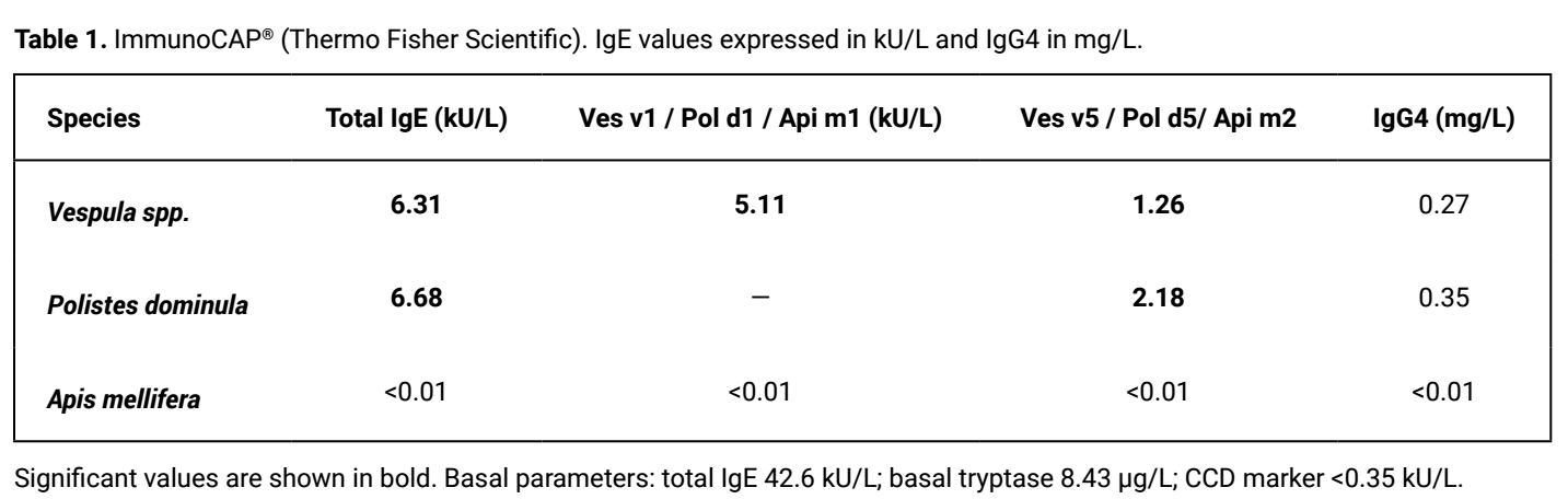
An allergologic study was performed using skin tests and laboratory studies. Prick and intradermal (ID) skin tests against Vespula spp. showed positivity in ID 0.1 mg/mL (7 × 5 mm) and against Polistes dominula in ID 0.1 mg/mL (5 × 5 mm), while skin tests for Apis mellifera were negative. Specific IgE determination was performed using ImmunoCAP® (Thermo Fisher Scientific) and a blot study for venoms using IMMULITE® 2000/2000XPi 3gAllergy™, which reported double sensitization to Vespula spp. and Polistes dominula, with the absence of sensitization against Apis mellifera and absence of sensitization to cross-reactive carbohydrate determinants (CCDs). Table 1
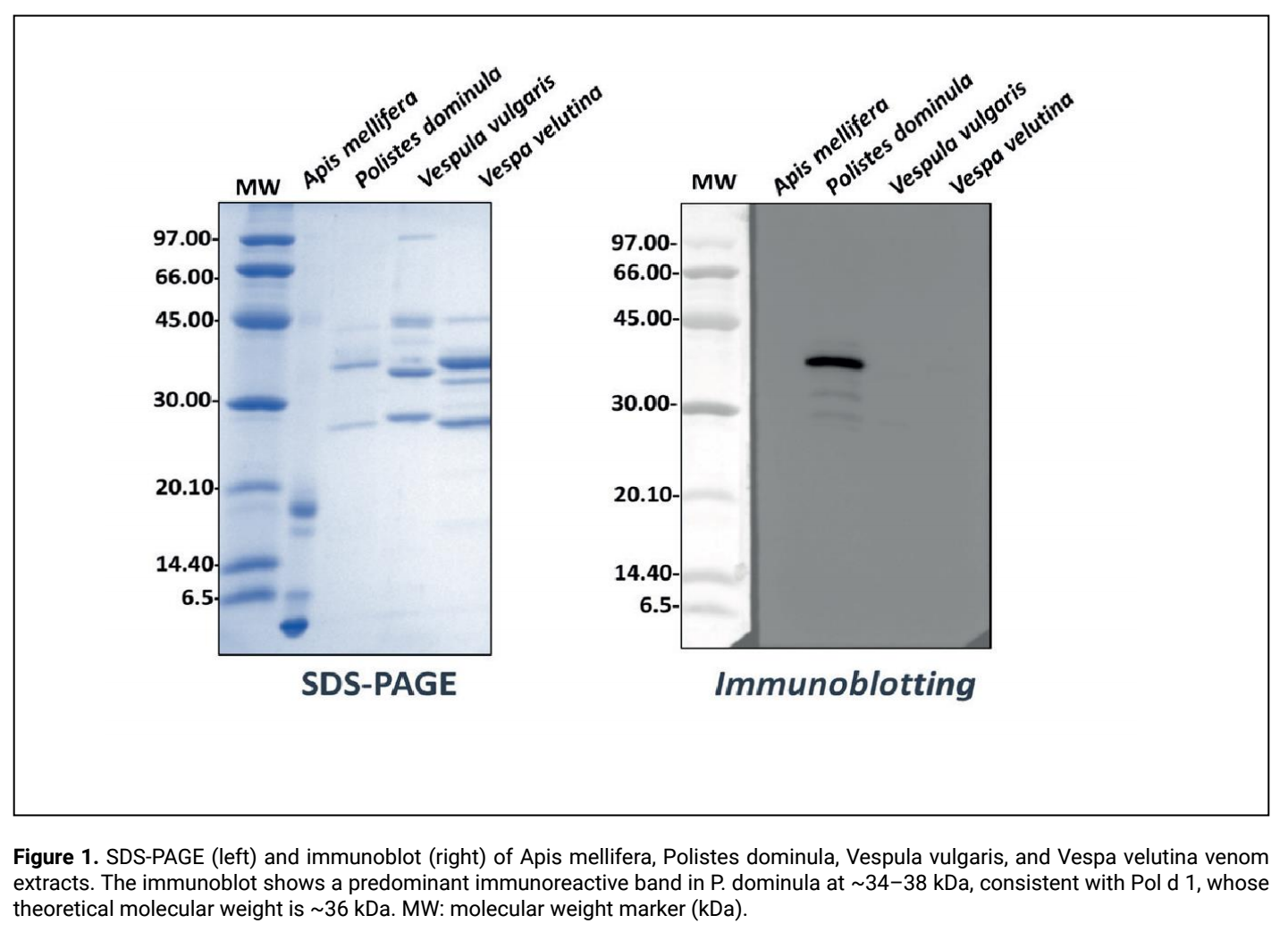
Additionally, an immunoblot was performed against venoms of the main Hymenoptera of allergologic relevance (Hymnox®/Venenox®, Roxall Medicina España S.A.), which revealed a predominant immunoreactive band in the Polistes dominula extract in the range of ~34–38 kDa, consistent with Pol d 1, whose theoretical molecular weight is ~36 kDa, which may appear increased under electrophoretic conditions due to glycosylation. Figure 1
A diagnosis of Polistes dominula allergy was established, and species-specific immunotherapy (SIT) was prescribed, with good tolerance and clinical response. Medical treatment in the event of a new sting was explained to the patient, and he was instructed on the use of an adrenaline autoinjector. After one year of starting SIT, he suffered a new accidental wasp sting in a public area without manifesting a systemic reaction, which supported the clinical efficacy of the treatment.
Due to a REMA score (Spanish Network on Mastocytosis) of +3 (high probability of clonal mast cell syndrome), a bone marrow study was performed. Cytology showed mast cell hyperplasia, mostly with atypical morphology (88% of the total). Flow cytometry detected 0.016% of aberrant mast cells, findings compatible with CMAS. The D816V mutation of the KIT gene, analyzed by digital PCR, was negative. Likewise, bone marrow densitometry and abdominal ultrasound were performed, with no pathological findings. Treatment with disodium cromoglycate (200 mg every 12 hours) was prescribed, with good subsequent clinical progression.5
DISCUSSION
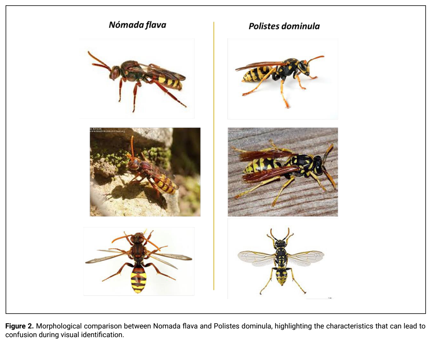
Nomada flava is a small cleptoparasitic bee belonging to the Apidae family, whose morphology can complicate visual identification and favor confusion with other hymenoptera of allergological relevance such as Apis mellifera or Polistes dominula. To date, there are no documented descriptions of allergic reactions to this species nor characterization of its allergens, which limits the availability of specific diagnostic tests. This situation forces reliance on integrating clinical history and available immunological techniques to identify the culprit insect.6–8
In our patient, the initial suspicion of a Nomada flava sting was inconsistent with the immunological findings, which showed predominant recognition against Pol d 1. This is the main allergen of Polistes dominula venom, and its detection is associated with clinically relevant sensitization.
In the ImmunoCAP® currently available in Spain, it is not possible to determine Pol d 1; therefore, complementary techniques (IMMULITE® and immunoblot) were used. These showed detectable concentrations of Pol d 1 (0.54 kU/L) and a predominant band in the expected range.
The predominant identification of Pol d 1, together with the absence of CCD-mediated cross-reactivity and negative results against Apis mellifera allergens, allowed for differentiation between serological double sensitization and clinically relevant Polistes dominula allergy. Therefore, it was considered more likely that the sting was caused by Polistes dominula, visually confused with Nomada flava due to their morphological similarity (Figure 2). The characteristics of the nest, the insect’s anatomy, and the immunological results support this hypothesis. The discovery of the stinger suggests the death of the insect after the sting, although this finding is not entirely specific. The visual difficulty in differentiating between species and the coexistence of double sensitization highlight the need for a comprehensive diagnostic approach that includes advanced techniques such as immunoblot, inhibition tests, or basophil activation tests to identify the correct species, thereby differentiating between primary sensitization and cross-reactivity.
The case presented in this study also shows that in patients with anaphylaxis due to Hymenoptera venom, especially in the most severe cases, clonal mast cell activation syndrome may coexist and go unnoticed, even with normal basal tryptase concentrations. Its detection is fundamental because it modifies clinical management, including the indication for prolonged or indefinite immunotherapy, carrying more than one adrenaline autoinjector, and the use of adjuvant treatments.
In our patient, the negative KIT D816V mutation, together with the absence of dense mast cell infiltration in the bone marrow, makes a diagnosis of indolent systemic mastocytosis less likely and supports the diagnosis of clonal mast cell activation syndrome, although both conditions can be part of a continuous spectrum and require longitudinal follow-up.
Among the limitations of the case presented is the impossibility of absolutely confirming the culprit species, since identification is based on indirect clinical and immunological findings. Likewise, as it is an isolated case, the extrapolation of the results should be performed with caution.
CONCLUSIONS
This case highlights the diagnostic complexity of Hymenoptera venom allergy and the importance of a multidimensional approach that integrates insect identification, assessment of cross-sensitizations, and the use of advanced diagnostic methods to ensure appropriate and effective immunotherapy. It also highlights the need to rule out clonal mast cell activation syndrome in patients with severe systemic reactions, due to its impact on prognosis and treatment.
Declaraciones
Author contributions
Each of the signing authors declares to have made substantial contributions that meet the four authorship criteria approved by the International Committee of Medical Journal Editors (ICMJE).
Conflict of interest
The authors declare no conflict of interest.
Funding
The authors declare that no funding was received for the conduct of this study.
Key references
Alfaya-Arias T, Soriano-Gómis V, Soto-Mera T, et al. Key Issues in Hymenoptera Venom Allergy: An Update. J Investig Allergol Clin Immunol 2017; 27 (1): 19-31. doi:10.18176/jiaci.0123Álvarez-Twose I, González-De-Olano D, Sánchez-Muñoz L, Matito A, et al. Validation of the REMA Score for Predicting Mast Cell Clonality and Systemic Mastocytosis in Patients with Systemic Mast Cell Activation Symptoms. Int Arch Allergy Immunol 2012; 157 (3): 275-280.Nomada flava Panzer, 1798: a new species of Cuckoo Bee for Spain and the Iberian Peninsula (Hymenoptera: Apoidea: Apidae), Álvarez M., Alvarez Fidalgo P., Baldock D. Boletín de la SEA, No. 65, 2019; 191-194.
Permissions
All figures and tables are original.
Permissions
All figures and tables are original.
Contribución de los autores
Cada uno de los autores firmantes declara haber realizado contribuciones sustanciales que cumplen los cuatro criterios de autoría aprobados por el Comité Internacional de Editores de Revistas Médicas (ICMJE).
Conflicto de intereses
Los autores declaran no tener conflictos de intereses.
Financiación
Los autores declaran que no se recibió financiación para la realización del presente estudio.
Referencias clave
Alfaya-Arias T, Soriano-Gómis V, Soto-Mera T, et al. Key Issues in Hymenoptera Venom Allergy: An Update. J Investig Allergol Clin Immunol 2017; 27 (1): 19-31. doi:10.18176/jiaci.0123Álvarez-Twose I, González-De-Olano D, Sánchez-Muñoz L, Matito A, et al. Validation of the REMA Score for Predicting Mast Cell Clonality and Systemic Mastocytosis in Patients with Systemic Mast Cell Activation Symptoms. Int Arch Allergy Immunol 2012; 157 (3): 275-280.Nomada flava Panzer, 1798: a new species of Cuckoo Bee for Spain and the Iberian Peninsula (Hymenoptera: Apoidea: Apidae), Álvarez M., Alvarez Fidalgo P., Baldock D. Boletín de la SEA, No. 65, 2019; 191-194.
Permisos
Todas las figuras y cuadros son originales.
Permisos
Todas las figuras y cuadros son originales.
REFERENCIAS
- 1.Popescu FD, Preda M, Antolín-Amérigo D, Rodríguez-Otero N, et al. Biomarkers for the Molecular Diagnosis of IgE-Mediated Hymenoptera Venom Allergy in Clinical Practice. Int J Mol Sci 2024; 26 (1): 270. doi:10.3390/ijms26010270
- 2.Alfaya-Arias T, Soriano-Gómis V, Soto-Mera T, et al. Key Issues in Hymenoptera Venom Allergy: An Update. J Investig Allergol Clin Immunol 2017; 27 (1): 19-31. doi:10.18176/jiaci.0123
- 3.Boggs NA, Tanasi I, Hartmann K, Zanotti R, et al. Mast Cell Disorders and Hymenoptera Venom-Triggered Anaphylaxis: Evaluation and Management. J Allergy Clin Immunol Pract 2025; 13 (1): 40-48. doi:10.1016/j.jaip.2024.08.034
- 4.Armisén M, Guspi R, Alfaya T, Cruz S, et al. Cross-Sectional Validation of a Quality of Life Questionnaire in Spanish for Patients Allergic to Hymenoptera Venom. J Investig Allergol Clin Immunol 2015; 25 (3): 176-182.
- 5.Álvarez-Twose I, González-De-Olano D, Sánchez-Muñoz L, Matito A, et al. Validation of the REMA Score for Predicting Mast Cell Clonality and Systemic Mastocytosis in Patients with Systemic Mast Cell Activation Symptoms. Int Arch Allergy Immunol 2012; 157 (3): 275-280.
- 6.Nomada flava Panzer, 1798: a new species of Cuckoo Bee for Spain and the Iberian Peninsula (Hymenoptera: Apoidea: Apidae), Álvarez M., Alvarez Fidalgo P., Baldock D. Boletín de la SEA, No. 65, 2019; 191-194.
- 7.Golden DBK, et al. Advances in the diagnosis of hymenoptera venom allergy. Curr Opin Allergy Clin Immunol 2020; 20 (4): 319-324.
- 8.Marquès L, Vega A, de la Roca F, et al. Molecular Diagnosis in Hymenoptera Allergy: Comparison of Euroline DPA-Dx and ImmunoCAP. Toxins (Basel) 2025; 17 (6): 310. doi:10.3390/toxins17060310.